Dentist Referral Form
André Hattingh
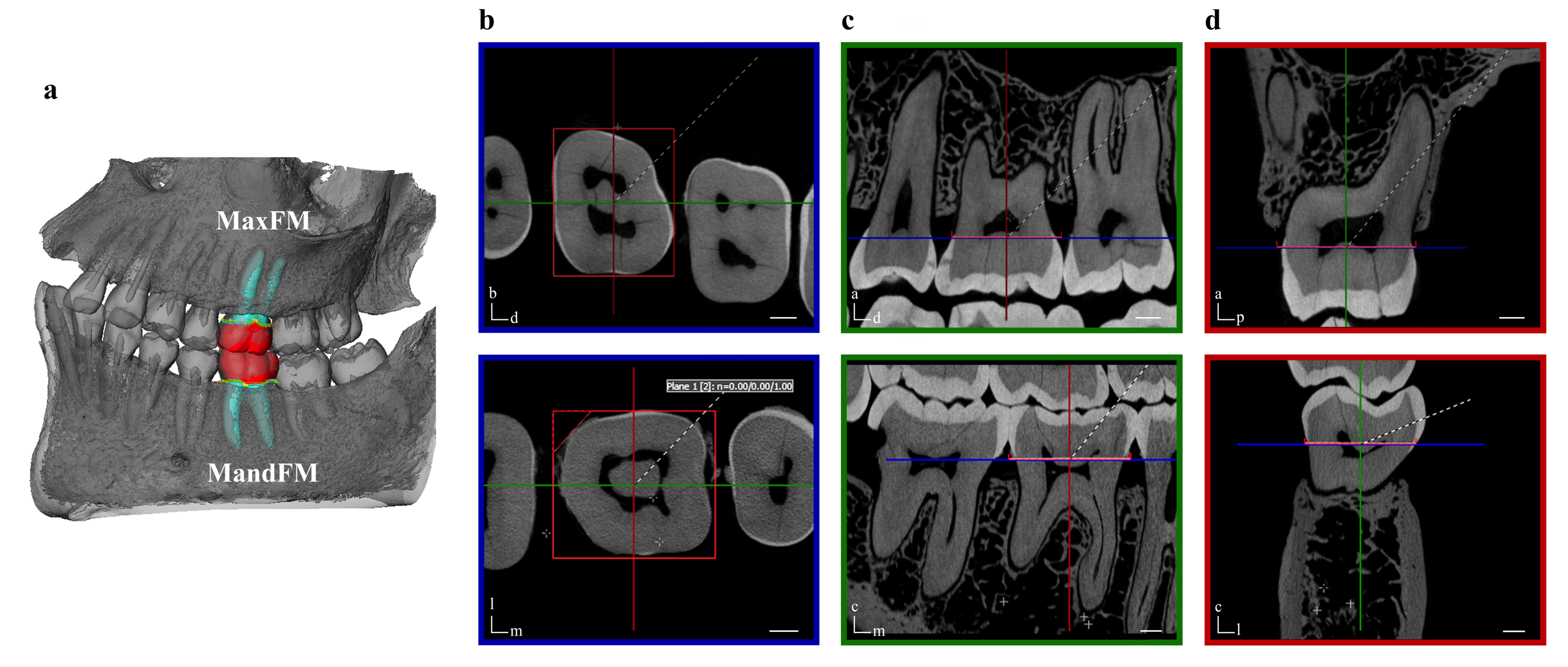
Micro CT study of molar sockets
Dento-alveolar measurements and histomorphometric parameters of maxillary and mandibular first molars, using micro-CT.
Theye CEG, Hattingh A, Cracknell TJ, Oettlé AC, Steyn M, Vandeweghe S.
Clin Implant Dent Relat Res. 2018 Aug;20(4):550-561. doi: 10.1111/cid.12616. Epub 2018 May 6.
PMID: 29732712
This peer reviewed article was published in collaboration with University of Pretoria, Medical University of South Africa, Wits University and Ghent University.
It is the first study of its kind that provides detailed information on human molar socket anatomy, which is vital to our understanding of the placement of dental implants in molar sockets.
Abstract
BACKGROUND:
Micro-CT is a high-resolution, non-invasive, and non-destructive imaging technique, currently acknowledged as a gold standard modality for assessing quantitatively and objectively dental morphology and bone microarchitecture parameters.
PURPOSE:
The aim of this study was to analyze critical dental and periodontal measurements characterizing the mandibular (MandFM) and maxillary (MaxFM) first molar architecture, as well as the corresponding bony socket, using micro-CT.
MATERIALS AND METHODS:
Thirty-eight human dried skulls (22-76 years) were scanned to enable the virtual analysis of 61 first molars. Depending on the type of measurement, the parameters were recorded on two-dimensional sections or directly on three-dimensional models. Tooth morphology was described by four aspects (e.g., tooth width, trunk length, root length, and root span), while the socket architecture was assessed by buccal plate thicknesses and bone density measurements.
RESULTS:
Minimum, maximum, and mean distances as well as cortical and trabecular bone densities were recorded in MandFM and MaxFM. It is noteworthy that the buccal plate thickness was found to be less than 1 mm in more than 55% of cases in MaxFM, whereas only in 20.8% of cases in MandFM (and even 0% at two sites). A wide range of bone densities was observed and the comparison between MandFM and MaxFM did not show a significant difference. Furthermore, cortical densities were negatively correlated with aging, while trabecular densities were not influenced.
CONCLUSIONS:
Using micro-CT, three-dimensional aspects of the human first molar morphology and microstructural parameters of the surrounding bone were evaluated in the mandible and in the maxilla. These comprehensive measurements and their correlation with aging may be of great importance for the use of immediate implant placement in molar extraction sockets and thus the potential long-term success of this treatment modality.